Practical ultrasound (fUS) marks a substantial leap in Brain-Machine User interface innovation, providing a less intrusive approach for exact control of electronic gadgets by analyzing brain activity.
Brain– device user interfaces (BMIs) are gadgets that can check out brain activity and equate that activity to manage an electronic gadget like a prosthetic arm or computer system cursor. They assure to allow individuals with paralysis to move prosthetic gadgets with their ideas.
Lots of BMIs need intrusive surgical treatments to implant electrodes into the brain in order to check out neural activity. Nevertheless, in 2021, Caltech scientists established a method to check out brain activity utilizing practical ultrasound (fUS), a much less intrusive strategy.
Practical Ultrasound: A Video Game Changer for BMIs
Now, a brand-new research study is a proof-of-concept that fUS innovation can be the basis for an “online” BMI– one that checks out brain activity, analyzes its significance with decoders set with artificial intelligence, and as a result manages a computer system that can precisely anticipate motion with extremely minimal hold-up time.
Ultrasound is utilized to image two-dimensional sheets of the brain, which can then be stacked together to produce a 3-D image. Credit: Thanks To W. Griggs
The research study was carried out in the Caltech labs of Richard Andersen, James G. Boswell Teacher of Neuroscience and director and management chair of the T&C Chen Brain– Maker User Interface Center; and Mikhail Shapiro, Max Delbrück Teacher of Chemical Engineering and Medical Engineering and Howard Hughes Medical Institute Detective. The work was a cooperation with the lab of Mickael Tanter, director of physics for medication at INSERM in Paris, France.
Benefits of Practical Ultrasound
” Practical ultrasound is an entirely brand-new technique to contribute to the tool kit of brain– device user interfaces that can help individuals with paralysis,” states Andersen. “It uses appealing choices of being less intrusive than brain implants and does not need continuous recalibration. This innovation was established as a really collective effort that might not be achieved by one laboratory alone.”
” In basic, all tools for determining brain activity have advantages and downsides,” states Sumner Norman, previous senior postdoctoral scholar research study partner at Caltech and a co-first author on the research study. “While electrodes can extremely exactly determine the activity of single nerve cells, they need implantation into the brain itself and are challenging to scale to more than a couple of little brain areas. Non-invasive strategies likewise include tradeoffs. Practical magnetic resonance imaging [ fMRI] offers whole-brain gain access to however is limited by minimal level of sensitivity and resolution. Portable approaches, like electroencephalography [EEG] are obstructed by bad signal quality and a failure to localize deep brain function.”
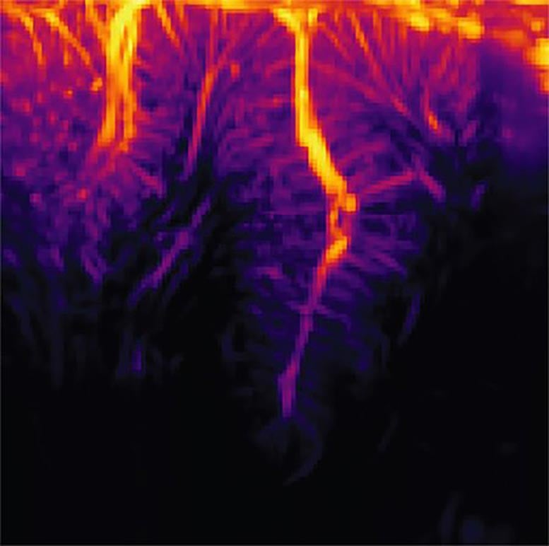
The vasculature of the posterior parietal cortex as determined by practical ultrasound neuroimaging. Credit: Thanks To W. Griggs
Ultrasound Imaging Explained
Ultrasound imaging works by producing pulses of high-frequency noise and determining how those sound vibrations echo throughout a compound, such as different tissues of the body. Acoustic waves take a trip at various speeds through these tissue types and show at the borders in between them. This strategy is typically utilized to take pictures of a fetus in utero, and for other diagnostic imaging.
Due to the fact that the skull itself is not permeable to acoustic wave, utilizing ultrasound for brain imaging needs a transparent “window” to be set up into the skull. “Significantly, ultrasound innovation does not require to be implanted into the brain itself,” states Whitney Griggs (PhD ’23), a co-first author on the research study. “This substantially minimizes the possibility for infection and leaves the brain tissue and its protective dura completely undamaged.”
” As nerve cells’ activity modifications, so does their usage of metabolic resources like oxygen,” states Norman. “Those resources are resupplied through the blood stream, which is the crucial to practical ultrasound.” In this research study, the scientists utilized ultrasound to determine modifications in blood circulation to particular brain areas. In the exact same method that the noise of an ambulance siren modifications in pitch as it moves closer and after that further away from you, red cell will increase the pitch of the shown ultrasound waves as they approach the source and reduce the pitch as they stream away. Determining this Doppler-effect phenomenon permitted the scientists to tape small modifications in the brain’s blood circulation to spatial areas simply 100 micrometers broad, about the width of a human hair. This allowed them to all at once determine the activity of small neural populations, some as little as simply 60 nerve cells, extensively throughout the brain.
Opening Motion: Assisting Paralyzed Individuals Usage Idea to Control Computers and Robotic Limbs
Ingenious Application in Non-Human Primates
The scientists utilized practical ultrasound to determine brain activity from the posterior parietal cortex (PAY PER CLICK) of non-human primates, an area that governs the preparation of motions and adds to their execution. The area has actually been studied by the Andersen laboratory for years utilizing other strategies.
The animals were taught 2 jobs, needing them to either strategy to move their hand to direct a cursor on a screen, or strategy to move their eyes to take a look at a particular part of the screen. They just required to believe about carrying out the job, not in fact move their eyes or hands, as the BMI checked out the preparation activity in their pay per click.
” I keep in mind how excellent it was when this sort of predictive decoding dealt with electrodes twenty years back, and it’s remarkable now to see it deal with a much less intrusive approach like ultrasound,” states Shapiro.
Promising Outcomes and Future Strategies
The ultrasound information was sent out in real-time to a decoder (formerly trained to decipher the significance of that information utilizing artificial intelligence), and consequently produced control signals to move a cursor to where the animal meant it to go. The BMI had the ability to effectively do this to 8 radial targets with mean mistakes of less than 40 degrees.
” It’s considerable that the strategy does not need the BMI to be recalibrated every day, unlike other BMIs,” states Griggs. “As an example, envision requiring to recalibrate your computer system mouse for approximately 15 minutes every day before usage.”
Next, the group prepares to study how BMIs based upon ultrasound innovation carry out in human beings, and to even more establish the fUS innovation to allow three-dimensional imaging for enhanced precision
The paper is entitled “Decoding motor strategies utilizing a closed-loop ultrasonic brain– device user interface” and was released in the journal Nature Neuroscience on November 30.
Recommendation: “Deciphering motor strategies utilizing a closed-loop ultrasonic brain– device user interface” by Whitney S. Griggs, Sumner L. Norman, Thomas Deffieux, Florian Segura, Bruno-Félix Osmanski, Geeling Chau, Vasileios Christopoulos, Charles Liu, Mickael Tanter, Mikhail G. Shapiro and Richard A. Andersen, 30 November 2023, Nature Neuroscience
DOI: 10.1038/ s41593-023-01500-7
Whitney Griggs (PhD ’23), UCLA– Caltech MD/PhD trainee, and Sumner Norman, previous postdoctoral scholar now of Forest Neurotech, are the research study’s very first authors. In addition to Griggs, Norman, and Andersen, Caltech coauthors are college student Geeling Chau and Vasileios Christopoulos, going to partner in biology and biological engineering. Other coauthors are Charles Liu of USC; and Mickael Tanter, Thomas Deffieux, and Florian Segura of INSERM in Paris, France. Financing was supplied by the National Eye Institute, a Josephine de Karman Fellowship, the UCLA-Caltech MSTP, the Della Martin Structure, the National Institute of Neurological Conditions and Stroke, the National Institutes of Health, the T&C Chen Brain-Machine User Interface Center, and the Boswell Structure.